UVA Health · New patients

Educational X-Ray Images

Posterior cervical fusion

Teaching films from Dr. Li’s surgery library. They are not a diagnosis. Your own studies should be read in clinic with the history and the exam.
Back to Posterior fusion

Posterior cervical fusion C2–T2
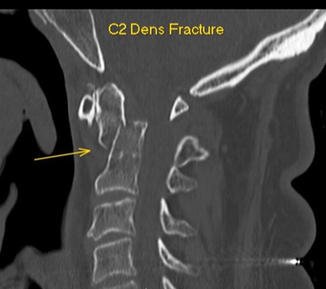
Sagittal CT of a C2 dens fracture
C2 dens fracture — preoperative sagittal CT
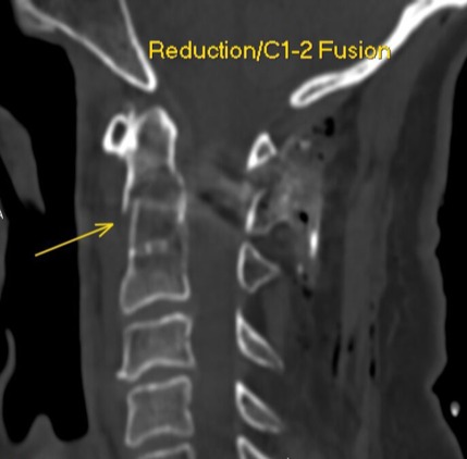
Sagittal CT after reduction and C1–2 posterior fusion
Reduction and C1–2 fusion — postoperative sagittal CT
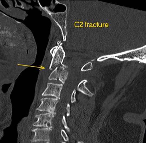
Sagittal CT of a C2 fracture
C2 fracture — preoperative sagittal CT
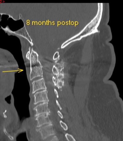
Sagittal CT eight months after C2 fracture treatment
C2 fracture — 8 months after surgery
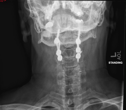
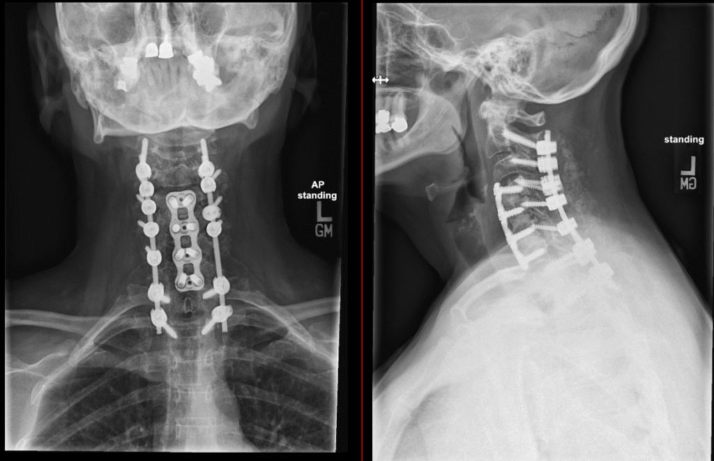
Standing AP x-ray after posterior cervical fusion
Posterior cervical fusion — standing AP
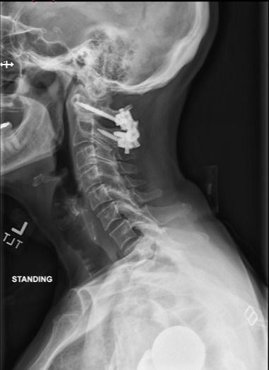
Standing lateral x-ray after posterior cervical fusion
Posterior cervical fusion — standing lateral
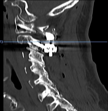
Postoperative sagittal CT after C1–2 fusion with screws
C1–2 fusion — postoperative sagittal CT
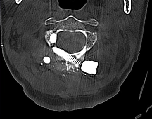
Postoperative axial CT after C1–2 fusion with screws
C1–2 fusion — postoperative axial CT
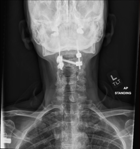
Standing AP x-ray after C1–2 posterior fusion
C1–2 fusion — standing AP
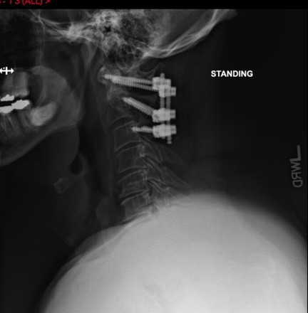
Standing lateral x-ray after C1–2 posterior fusion
C1–2 fusion — standing lateral

Images are de-identified educational examples. They do not show every variation of this operation, and they are not a promise of a particular result.

Appointments

Ready to take the next step toward recovery?

New patients are welcome. Call the spine clinic or send a note — the team will help you find the right visit.